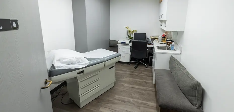
Endoscopic Ultrasound
in Los Angeles
Understanding the Endoscopic Ultrasound Procedure

An endoscopic ultrasound (EUS) combines the visual precision of endoscopy with the structural depth of ultrasound imaging, providing Dr. Sikavi with a uniquely detailed view of the gastrointestinal tract and the surrounding organs. While a standard endoscopy examines the surface lining of the GI tract, EUS goes further, producing high-resolution images of the deeper layers of the GI wall and nearby structures, including the pancreas, bile ducts, gallbladder, liver, and lymph nodes.
For patients whose symptoms remain unexplained after standard testing, EUS frequently provides the clarity needed to move forward with a confident diagnosis and treatment plan.
Should You Schedule an Endoscopic Ultrasound?

Dr. Sikavi develops a targeted diagnostic plan for each patient based on prior test results, imaging findings, and clinical presentation, not a one-size-fits-all protocol.
What an Endoscopic Ultrasound Diagnoses
01
Pancreatic Conditions
EUS is considered the most sensitive imaging modality for evaluating the pancreas. It can identify small tumors, cysts, and inflammatory changes that other imaging methods routinely miss, and allows for direct tissue sampling when needed.
02
GI Cancer Staging
For cancers of the esophagus, stomach, rectum, and pancreas, EUS provides precise information about tumor depth and lymph node involvement, which is critical for determining the most appropriate treatment approach.
03
Bile Duct and Gallbladder Disease
Stones lodged in the bile duct, biliary strictures, and other abnormalities in the biliary system are clearly visualized with EUS, often with greater accuracy than standard ultrasound or CT.
04
Submucosal Lesions
Growths that originate beneath the surface lining of the GI tract are difficult to characterize with standard endoscopy alone. EUS defines their size, layer of origin, and tissue characteristics to guide next steps.
05
Enlarged Lymph Nodes
When lymph nodes near the GI tract appear abnormal on imaging, EUS allows Dr. Sikavi to visualize and biopsy them directly, providing a tissue diagnosis without the need for surgery.
06
Chronic Pancreatitis
EUS detects structural changes in the pancreatic tissue and ductal system that are characteristic of chronic pancreatitis, even in early stages when other imaging may appear normal.
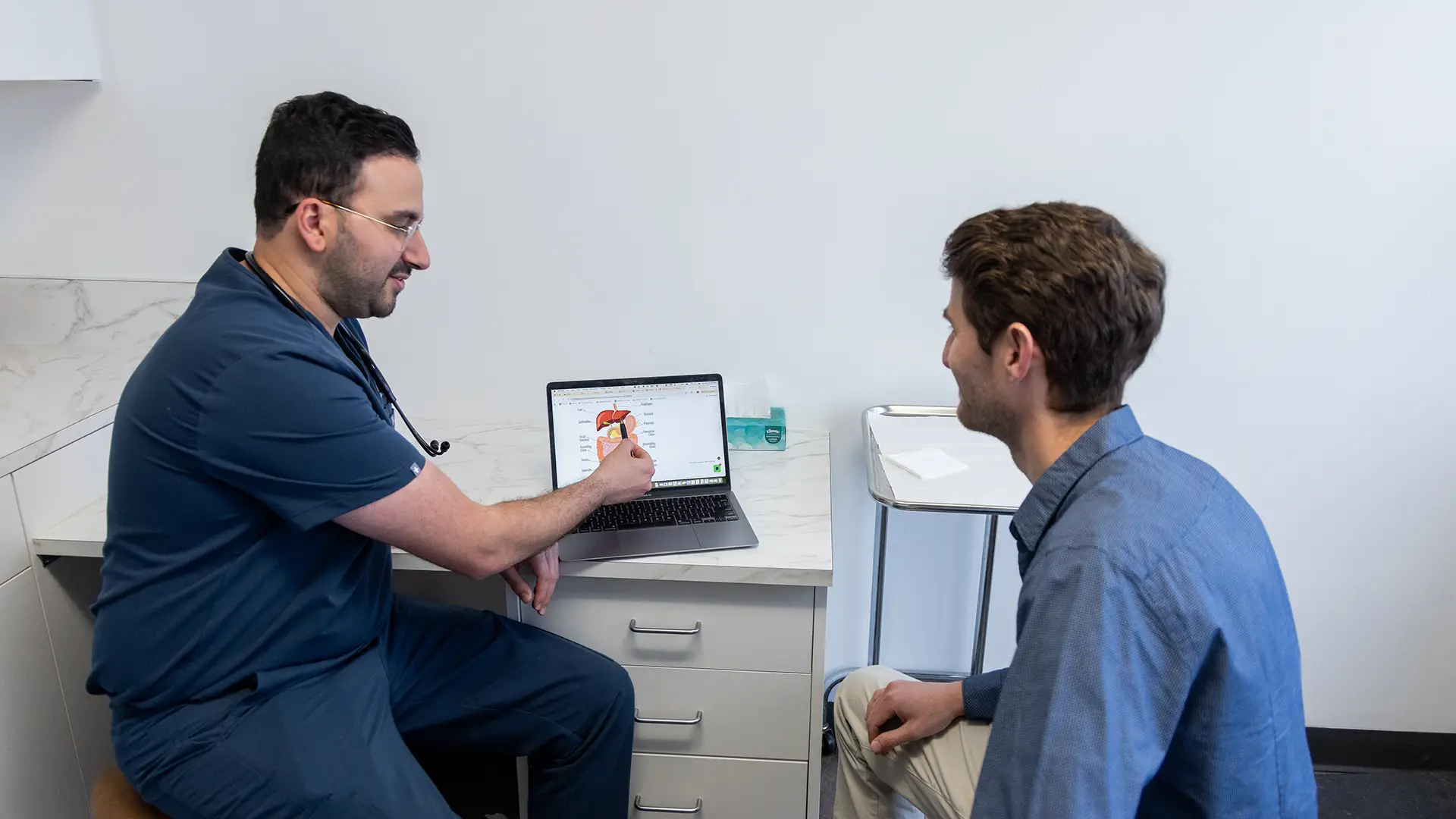
What Happens During an Endoscopic Ultrasound?


Frequently Asked Questions About Endoscopic Ultrasound
Why Patients Choose Innovative GI for Endoscopic Ultrasound in Los Angeles
Endoscopic ultrasound is a technically demanding procedure that requires specialized fellowship training and a high level of procedural experience. Not all gastroenterologists perform EUS, and fewer still bring the investigative precision that complex cases require.
Our Los Angeles gastroenterology practice, located directly across from Cedars-Sinai Medical Center in the heart of West Hollywood and Beverly Grove, was designed to feel different from a typical GI clinic:
- Advanced EUS equipment for high-resolution deep-layer imaging
- Integrated fine needle aspiration and biopsy capability for same-session tissue sampling
- Sedation-assisted procedures for a comfortable, stress-free experience
- A focused, concierge-level environment where Dr. Sikavi gives you his full, undivided attention
- Convenient access from Beverly Hills, West Hollywood, Mid-Wilshire, and the broader Westside